Your sensitivity to light and color depends on specific anatomical factors that vary between individuals. If you’ve got light-colored irises, you have less melanin to block incoming UV and visible light. Your retinal pigment epithelium concentration also affects how much light reaches your photoreceptors. Differences in cone density alter how you process color wavelengths, while stress-induced pupil dilation and age-related muscle decline further compound photosensitivity. Understanding each variable can help you identify your triggers and the right protective interventions.
How Iris Color Affects Your Sensitivity to Light
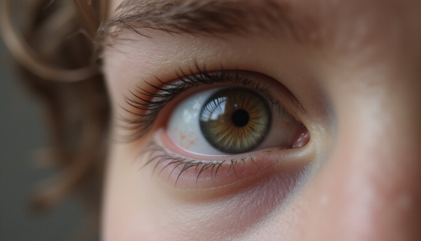
If you’ve ever squinted painfully under fluorescent lights while the person beside you seemed unbothered, your iris color likely plays a role. Light-colored irises, blue, green, grey, contain fewer melanocytes with loosely arranged stromal fibers. This reduced melanin density permits greater ultraviolet and visible light penetration through the iris stroma, increasing retinal exposure. Your thin iris functions like a lace curtain rather than heavy brocade, amplifying intraocular straylight. Research measuring 853 participants confirms light-blue irises produce notably higher straylight values (1.14 log) compared to brown irises (1.06 log).
This heightened sensitivity to light and color anxiety around bright environments isn’t psychological, it’s anatomical. You’ll experience pronounced pupillary constriction reflexes, reduced contrast sensitivity, and disability glare during routine activities like night driving. To minimize discomfort and protect against long-term damage, light-eyed individuals should prioritize wearing UV-protective sunglasses, wide-brimmed hats, and seeking shade during peak sunlight hours.
Why Your Retinal Pigment Layer May Not Filter Enough Light
Your retinal pigment epithelium‘s melanin density isn’t uniform across all individuals, it varies based on genetic factors, meaning your RPE may provide substantially less baseline photoprotection than someone else’s. If you have lighter eye pigmentation, your RPE likely contains lower melanin concentrations, reducing its capacity to absorb harmful wavelengths before they reach vulnerable photoreceptors. Because your body can’t synthesize new RPE melanin after birth, any baseline deficiency in this pigment layer represents a permanent limitation in your eyes’ intrinsic light-filtering defense. As RPE cells accumulate A2E lipofuscin fluorophores over time, this already-limited protection becomes further compromised, making them increasingly susceptible to blue light-induced damage.
RPE Density Varies Individually
Behind every photoreceptor in your eye sits a single layer of cells called the retinal pigment epithelium (RPE), a cellular shield that absorbs stray light, recycles visual pigments, and maintains the metabolic health of the cones and rods it supports.
Your foveal RPE density ranges from 8,700 to 14,800 cells/mm², substantial inter-individual variation that directly affects light filtration capacity. Critically, cone density and RPE density vary independently, producing cone-to-RPE ratios spanning 8:1 to 21:1 at the fovea. If your ratio skews high, each RPE cell bears greater metabolic load, potentially compromising absorption efficiency. This mismatch can heighten your sensitivity to colors stress response under ordinary illumination. RPE density also declines approximately 0.23% annually, with macular cell loss accelerating after age sixty, compounding filtration deficits progressively across your lifespan. Research on cadaver eyes confirms that RPE cells in the far periphery show increasing area and eccentricity with age, meaning the disorganization compounds with distance from the central retina outward.
Light-Eye Pigment Deficiency
Because melanin concentration in your iris stroma dictates how much light passes through to the retina, lighter eye colors, blue, gray, and green, function with a measurably thinner optical filter than brown or black irises. Your retinal pigment epithelium compounds this deficit when its melanin density falls below protective thresholds, allowing excessive photon stimulation of rod and cone photoreceptors.
This cumulative pigment deficiency, spanning both iris and RPE, creates pronounced photophobia. You’ll notice discomfort in environments others tolerate easily. In severe cases, such as ocular or oculocutaneous albinism, near-total melanin absence produces debilitating light intolerance that impairs reading, screen use, and outdoor activity. The resulting hyperactivation of visual pathways can trigger light sensitivity anxiety symptoms, where anticipated exposure to bright environments generates physiological stress responses beyond standard photophobic discomfort.
Why Your Eyes Literally See Different Colors Than Someone Else’s
Though you and a friend may look at the same red apple under identical lighting, your retinas don’t process that experience the same way. Your eyes contain approximately six million cone cells, yet cone density varies considerably between individuals. Three distinct cone types, short (445 nm), medium (535 nm), and long (565 nm), detect different wavelengths, but their distribution across your retina differs from anyone else’s.
Despite these anatomical variations, your visual cortex standardizes incoming signals. A 2005 University of Rochester study confirmed that individuals perceive basic colors similarly regardless of cone count differences. However, genetic mutations like tetrachromacy introduce a fourth cone type, potentially expanding color discrimination. Understanding your unique cone configuration matters because heightened color sensitivity mental health impacts include overstimulation, anxiety, and sensory fatigue in environments with intense chromatic exposure.
How Stress and Mood Can Make Your Eyes More Light-Sensitive
When stress activates your sympathetic nervous system, your pupils dilate as part of the fight-or-flight response, allowing more light to flood the retina and intensifying photosensitivity. Elevated cortisol and adrenaline heighten sensory perception, making standard illumination feel overwhelming. This explains how stress and mood can make your eyes more light-sensitive through measurable neurochemical pathways. The impact of sensory overload on anxiety can be profound, as individuals may find themselves overwhelmed by their environment. This heightened state of alertness can lead to increased feelings of unease and tension.
Light-sensitive retinal ganglion cells transmit intensity signals to your prefrontal cortex and thalamus, directly modulating affective processing. When these mood-regulating pathways are chronically activated, even normal ambient light triggers irritability and emotional dysregulation. Simultaneously, visual sensitivity causes anxiety through a cyclical mechanism, photophobia amplifies distress, which further dilates pupils and lowers your discomfort threshold. Prolonged blue light exposure compounds this by suppressing melatonin and elevating cortisol, reinforcing the feedback loop between photosensitivity and psychological distress.
How Aging Increases Light and Color Sensitivity
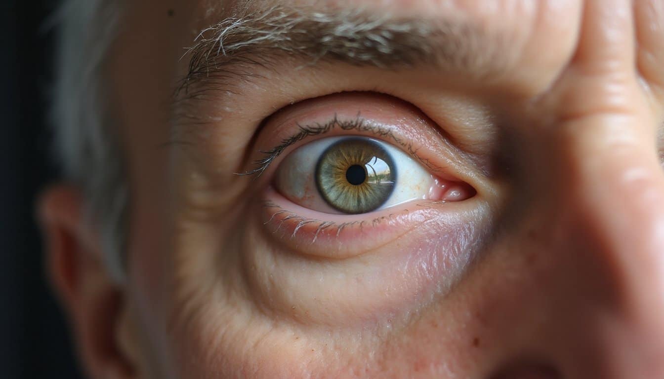
As you age, your eye’s ciliary and iris muscles weaken, reducing your pupil’s ability to regulate incoming light and causing heightened glare sensitivity. Your retina simultaneously loses cone and rod photoreceptors, diminishing your capacity to detect color wavelengths, particularly in the blue spectrum, and reducing overall visual acuity under low-contrast conditions. This dual decline impairs your brightness adaptation mechanism, making shifts between light and dark environments slower and more visually disorienting.
Weakening Eye Muscle Response
The iris sphincter muscles govern automatic pupil constriction in response to bright light, and their progressive weakening with age undermines this protective mechanism. When your iris sphincter muscles deteriorate, they can’t contract effectively, leaving your pupils abnormally dilated under bright conditions. This weakening eye muscle response permits excessive photic input through the pupillary aperture, directly intensifying light sensitivity.
As your pupils lose contractile efficiency, you’ll experience heightened discomfort in environments you previously tolerated. The mechanical failure in your iris musculature eliminates your eye’s primary defense against luminous overexposure. This dysfunction often triggers photophobia anxiety, as unpredictable light encounters become sources of distress. You may find yourself avoiding well-lit spaces or squinting reflexively, compensating for what your iris sphincter muscles can no longer regulate autonomously.
Declining Retinal Cell Sensitivity
Although your retina’s photoreceptor layer thins at an average rate of 0.143 micrometers per year, measured most prominently at the foveal minimum, this structural degradation doesn’t heighten your sensitivity to light and color. Declining retinal cell sensitivity actually diminishes your visual function progressively.
Key anatomical changes include:
- Rod photoreceptor selective loss: Rods deteriorate faster than cones, severely compromising your scotopic vision while relatively preserving photopic capacity
- RPE dysfunction: Your retinal pigment epithelium loses cellular density in the central macula, impairing light-sensitive molecule recycling and neuroprotective functions
- Contrast sensitivity reduction: Linear decline occurs from ages 20 to 74, with parafoveal thresholds correlating more strongly with age advancement
Physical inactivity and smoking accelerate photoreceptor layer thinning, compounding your age-related structural losses beyond typical degradation patterns.
Impaired Brightness Adaptation
Because your eyes’ adaptive mechanisms deteriorate with age, brightness shifts that once felt seamless now produce prolonged visual discomfort and heightened sensitivity. Your pupils constrict less efficiently, reducing retinal illumination control, while choroidal thinning limits retinoid cycling essential for photoreceptor recovery. These compounding deficits create impaired brightness adaptation that worsens progressively beyond age fifty.
Your dark adaptation curve shifts rightward, extending recovery time constants as rod-mediated scotopic sensitivity declines across the parafovea and macula. Simultaneously, cataracts reduce light transmission, further disrupting photopic-scotopic changes. Neural degradation within both magnocellular and parvocellular pathways amplifies visual overstimulation sensitivity, particularly at high spatial frequencies. Your contrast sensitivity deteriorates more notably in the magnocellular pathway, meaning rapid luminance changes trigger disproportionate discomfort. These concurrent retinal, lenticular, and neural changes produce measurable adaptation failure.
Fluorescent Lights, Screens, and Other Triggers That Worsen Sensitivity
Fluorescent lighting ranks among the most common environmental triggers that markedly intensify visual sensitivity, largely due to its unique spectral composition and low-frequency flicker. Your neural visual processing centers detect these imperceptible pulse transmissions, activating neurological responses that manifest as headaches, migraines, and ocular strain. This sensory sensitivity light exposure interaction affects even individuals without pre-existing conditions.
Common symptom manifestations include:
- Ocular disturbances: excessive lacrimation, involuntary blink reflex, erythema, xerophthalmia, and accommodative dysfunction
- Neurological responses: fatigue, vertigo, nausea, and cognitive performance degradation
- Visual disruptions: diplopia, blurred acuity, and difficulty adjusting to luminance intensity changes
Conditions such as autism spectrum disorder, photophobia, traumatic brain injury, and Irlen syndrome markedly heighten your susceptibility, making fluorescent environments particularly problematic for daily functioning. Cognitive behavioral therapy techniques can be beneficial in managing the symptoms associated with these conditions. These strategies help individuals develop coping mechanisms and improve their daily interactions.
Practical Ways to Protect Your Eyes From Light Sensitivity
When your visual system reacts intensely to environmental light stimuli, targeted protective interventions can substantially reduce ocular discomfort and neurological symptom burden. Select sunglasses blocking 100% UVA and UVB radiation with wrap-around frames to eliminate peripheral light infiltration. FL-41 rose-tinted lenses filter blue-green wavelengths linked to photophobia and migraine activation. Natural light can play a significant role in creating a tranquil environment. The calming effects of natural light can improve mood and focus, making it an ideal addition to both workspaces and homes.
Indoors, install dimmer switches and replace fluorescent sources with warm-toned LEDs to minimize visual sensitivity anxiety triggers. Apply anti-glare coatings to corrective eyewear and implement the 20-20-20 rule during screen exposure. Adjust display settings to dark mode, reducing retinal strain from artificial luminance. Layer window treatments, curtains paired with blinds, for precise ambient light regulation. Preservative-free lubricating drops maintain corneal surface integrity, supporting sustained ocular comfort throughout daily activities.
Call Now and Get the Help You Need
Anxiety has a way of making everyday life feel heavier than it should but real relief is within reach when you have the right people beside you. At Villa Healing Center, we provide Anxiety Treatment built around your needs to help you find lasting peace. Serving individuals throughout Los Angeles County, our compassionate team is ready when you are. Call (888) 669-0661 today and take the first step toward healing.
Frequently Asked Questions
Can Certain Foods or Supplements Help Reduce Sensitivity to Colors and Light?
Yes, you can support your visual system through targeted nutrition. Beta-carotene from carrots and sweet potatoes strengthens your retinal adaptation, while lutein and zeaxanthin from dark leafy greens filter blue light at the macular level. You’ll also benefit from green tea’s EGCGs, which neutralize UV-induced free radicals in dermal cells. Omega-3 fatty acids and anthocyanin-rich foods like blackberries further protect your retinas against photosensitive oxidative stress responses.
Is Light Sensitivity a Sign of an Underlying Neurological Condition?
Yes, light sensitivity can indicate an underlying neurological condition. You’ll find it most commonly associated with migraines, where it’s the second most frequent symptom. It also presents in traumatic brain injury, blepharospasm, meningitis, and encephalitis. Your intrinsically photosensitive retinal ganglion cells activate trigeminal brainstem neurons and thalamic pain pathways, driving photophobia. If you’re experiencing persistent sensitivity alongside fever, neck stiffness, or altered consciousness, you should seek urgent neurological evaluation.
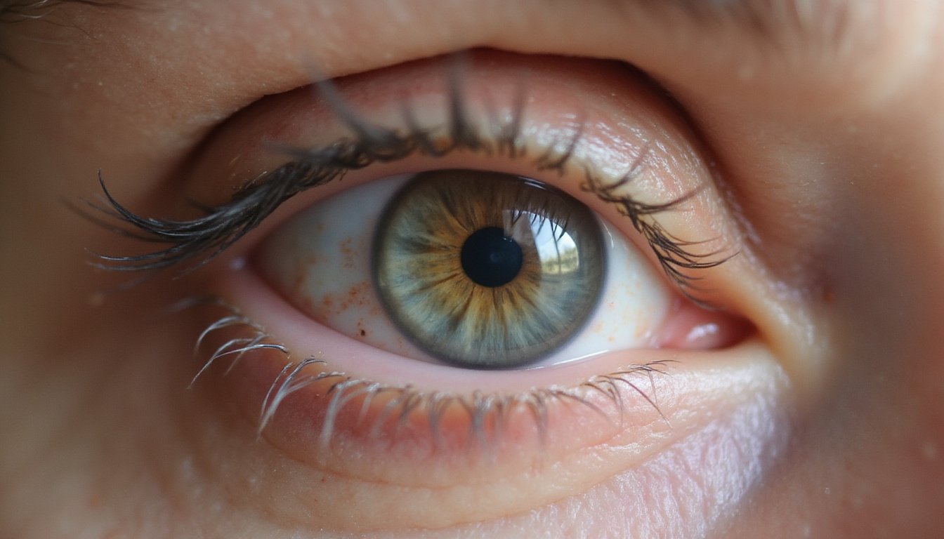
Are Children More Sensitive to Light and Color Than Adults?
Yes, children typically exhibit greater photosensitivity than adults. Their developing nervous systems demonstrate heightened responsiveness to flickering lights and specific wavelengths, particularly blue-green spectra. You’ll notice children can’t easily verbalize their discomfort, they’ll squint, cover their eyes, or show irritability instead. If your child has sensory processing differences, ADHD, or autism spectrum disorder, they’re especially vulnerable. Fluorescent classroom lighting and screen exposure can worsen their photophobic responses considerably.
Can Light Sensitivity Be Permanently Cured With Surgery or Medical Treatment?
Refractive surgeries like LASIK and LASEK can permanently reduce your photophobia by correcting corneal irregularities and refractive strain. You’ll likely experience temporary light sensitivity post-operatively, but it typically resolves within three months as your corneal epithelium regenerates. However, surgery won’t guarantee a complete cure. If you’ve had retinal detachment, you’ll retain residual light sensitivity loss regardless of surgical method. Chronic dry eye or migraine-related photophobia may also cause persistent symptoms.
Does Screen Time During Childhood Increase Lifelong Light and Color Sensitivity?
Research suggests childhood screen time can alter your color perception development. Studies show moderate screen exposure at age six correlates with modified tritan-axis discrimination and achromatic contrast sensitivity thresholds by adolescence. Blue light from digital devices stimulates your retinal photoreceptor cells responsible for short-wavelength detection, potentially enhancing chromatic sensitivity. However, you should note that cumulative blue light’s long-term effects on retinal tissue and persistent photosensitivity pathways remain clinically undetermined, requiring further longitudinal investigation.